Molecular Mechanism of Tumor Suppression by C/EBPβ 3 UTR
Source:
Time: 2011-02-16
A team of researchers, led by LIU Dinggan at the Institute of Biochemistry and Cell Biology, Shanghai Institutes for Biological Sciences, CAS, found the molecular mechanism of the tumor suppression by the 3’untranslated region (3’UTR) of the mRNA of transcription factor C/EBPβ.
The functions of the 3’UTR as an integral part of a eukaryotic mRNA is well known. However, the 3’UTR which is isolated from mRNA and exists within eukaryotic cells, i.e. independent 3’UTR RNA, has not been well studied up to now, and even its existence is in controversy.
The C/EBPβ 3’UTR is an independent 3’UTR RNA first (1991) found by LIU Dinggan’s team. It is a tumor suppressor. Recently, this team found that the tumor suppression effect was exerted by the interactions between C/EBPβ 3’UTR and protein kinase Cε, the oncogene responsible for the malignancy in the hepatoma cell line under study. PhD student WANG Ying et al. disclosed that the phosphorylation of some proteins, including keratin 18, was reduced by the transfection of C/EBPβ 3’UTR RNA; the reduction in keratin phosphorylation was due to the inhibition of protein kinase Cε activity; and it was just this protein kinase (PK) variant which was the chief cause of the malignant phenotype of the said tumor cells. Then they showed that, in in vitro tests, C/EBPβ 3’UTR RNA did inhibit the phosphorylaiton by PKCε; and, in vivo, C/EBPβ 3’UTR RNA formed an RNA-protein complex with PKCε and keratin 18 molecules and therefore inhibited the activity of the PKCε. This is why the C/EBPβ 3’UTR suppresses malignancy of tumor cells.
This research entitled "Tumor suppression by RNA from C/EBPβ 3’UTR by the inhibition of PKCε activity" was published in PLoS ONE on January 24, 2011. This work was supported by grants from the National Natural Science Foundation of China. (SIBCB)
AUTHOR CONTACT:
LIU Dinggan
Institute of Biochemistry and Cell Biology, Shanghai Institutes for Biological Sciences, Chinese Academy of Sciences, Shanghai 200031, China
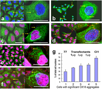
C/EBPβ 3′UTR RNA binds CK18 and alters its intracellular organization in SMMC-7721 and Cl1 cells. Confocal micrographs of the cells immunostained with a fluorescein-labeled antibody against CK18 (green), and of the cells immunostained as above and probed with a fluorescent-labeled molecular beacon specific to C/EBPβ 3′UTR RNA (red dots). (a) SMMC-7721 cells. Arrows indicate the thin CK18 network in the cytoplasm. (b) Cl1 cells. Arrows indicate the aggregates and bundles of CK18. (c) SMMC-7721 cells probed with the molecular beacon for C/EBPβ 3′UTR RNA. Arrows indicate the molecular beacon binding with C/EBPβ 3′UTR RNA on the CK18 filaments. (d) Cl1 cells probed with the same molecular beacon. Note the greater amount of red fluorescence from the molecular beacon than in SMMC-7721, and the positioning of the fluorescence both around the CK18 filaments and even on the CK18 aggregates (the orange arrows in the larger amplification). (e) SMMC-7721 cells transfected with 1 µg/well of C/EBPβ 3′UTR RNA and probed with the same molecular beacon. Arrows indicate the fluorescence of the molecular beacon on the CK18. (f) SMMC-7721 cells transfected with 5 µg/well of C/EBPβ 3′UTR RNA and probed with the same molecular beacon. Arrows indicate the fluorescence of the molecular beacon on the CK18. Bars, 10 µm. (g) Percentage amounts of cells with significant CK18 aggregates in total cell populations of SMMC-7721, Cl1 and SMMC-7721 transfected with varying amounts of C/EBP 3′UTR RNA. Cells were grown in 24-well plates. 1, SMMC-7721. 2-4, SMMC-7721 transfected with 1, 3, 5 µg/well of C/EBP 3′UTR RNA respectively. 5, Cl1.
 Appendix: