The 35-kDa Fragment of TDP-43 Triggers Cytoplasmic Inclusion Formation and Altered RNA Processing
Source:
Time: 2011-04-11
TAR DNA binding protein of 43 kDa (TDP-43) is a nuclear factor functioning in RNA processing. It is also a major deposited protein in amyotrophic lateral sclerosis (ALS) and frontotemporal lobar degeneration with ubiquitin (FTLD-U). Under pathological conditions, TDP-43 is fragmented into two major C-terminal forms: ~35 kDa and ~25 kDa in the inclusions. Recently, a team of researchers, led by HU Hongyu, at the Institute of Biochemistry and Cell Biology, Shanghai Institutes for Biological Sciences, CAS, demonstrated that the 35-kDa fragment of TDP-43 plays an important role in the formation of inclusion bodies and alters the RNA processing in cells.
TDP-43 is comprised of an N-terminal random coil, two RNA recognition motifs (RRMs), and a C-terminal glycine rich region (GRR). To understand the mechanism underlying the inclusion formation and possibly functional alteration, CHE Meixia, a student in Dr. HU’s lab, studied some TDP-43 fragments and their effects on RNA processing in cells. The results show that the 35-kDa fragment of TDP-43 (namely TDP-35, residues 90–414) is capable of forming cytoplasmic inclusion bodies and altering pre-mRNA splicing. The inclusions formed by TDP-35 can also recruit full-length TDP-43 to cytoplasmic deposition from functionally nuclear localization. The in vitro studies demonstrated that TDP-35, rather than TDP-43 and TDP-25, is prone to aggregation, and it further serves as a seed to facilitate aggregation of full-length TDP-43. This suggests that fragmentation of TDP-43 leads to cellular redistribution, inclusion body formation, and altered RNA processing, which are implicated in the molecular pathogenesis of ALS and FTLD.
This work entitled “Aggregation of the 35-kDa fragment of TDP-43 causes formation of cytoplasmic inclusions and alteration of RNA processing” was published online in The FASEB Journal on Mar 30th, 2011.
This work was supported by the grants from the Chinese Academy of Sciences, the Ministry of Science and Technology, and the National Natural Science Foundation of China. (SIBCB)
AUTHOR CONTACT:
HU Hongyu
Institute of Biochemistry and Cell Biology, Shanghai Institutes for Biological Sciences, Chinese Academy of Sciences, Shanghai, China
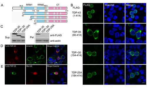
Formation of cytoplasmic inclusion bodies by exogenous TDP-43 fragments. A) Schematic representation of the domain architecture of TDP-43 and its truncations. NLS, nuclear localization signal; NES, nuclear export signal; RRM, RNA recognition motif. B) Localization and inclusion body formation of TDP-43 and its fragments. FLAG-tagged TDP-43 and its fragments were overexpressed in HEK 293T cells. The FLAG-tagged proteins are green, and the nuclei are stained with Hoechst (blue). C) Detection of the exogenous proteins in soluble and insoluble fractions. The cell lysates from HEK 293T cells overexpressing FLAG-tagged TDP-35, TDP-30 or TDP-25A were fractionated into supernatant (S) and pellet (P), and subjected to SDS-PAGE (12% gel), followed by immunoblotting with an anti-FLAG antibody. Both TDP-30 and TDP-25A are in the soluble fraction, whereas TDP-35 is in both fractions. D) Co-localization of inclusion bodies formed by TDP-35 with vimentin in HEK 293T cells. FLAG-TDP-35 is red and the endogenous vimentin is green. E) Co-localization of inclusion bodies formed by TDP-35 with ubiquitin in HEK 293T cells. FLAG-TDP-35 is green and Myc-ubiquitin is red. Scale bar, 10 μm. (Image provided by Dr. HU Hongyu)
 Appendix: