Virus infection triggers innate immune response and induces interferon and proinflammatory cytokines to restrict virus proliferation in higher organisms. Antiviral innate immune response is initiated with the detection of viral nucleic acids by pattern recognition receptors such as RIG-I. RIG-I binds to viral RNA and then activates MAVS in the presence of K63-linked polyubiquitin chains. MAVS (also known as IPS-1, VISA and CARDIF) is a mitochondrial protein and plays an essential role in antiviral innate immune signaling.
Recently, the lab led by Prof. HOU Fajian from the Institute of Biochemistry and Cell Biology, Shanghai Institutes for Biological Sciences, Chinese Academy of Sciences, reported that an autoinhibitory mechanism modulates MAVS activity in antiviral innate immune response.
Upon viral infection, RIG-I induces MAVS to form prion-like filaments. Consequently, MAVS becomes active to stimulate downstream signaling effectors, such as IKK and TBK1. Prion-like filament formation of MAVS does not involve protein upregulation or post-translational modification, raising an important question on how MAVS activity is regulated during the process. In this study, graduate students SHI Yuheng, YUAN Bofeng and postdoctoral fellow QI Nan, as leading authors, identified three functionally distinct active regions of MAVS for the activation of its downstream effectors through recruiting preferential TNF receptor-associated factors. Furthermore, they discovered that two of these regions are inhibited by their respective adjacent regions in the absence of upstream signal and are activated with the formation of MAVS filament on stimulation. Collectively, their findings uncovered an autoinhibitory mechanism that regulates these active regions in unstimulated MAVS, thus ensuring a prompt responsiveness of MAVS to virus infection.
Their work entitled “An autoinhibitory mechanism modulates MAVS activity in antiviral innate immune response” was published online in Nature Communications on July 17th, 2015. This study was supported by grants from Program, Shanghai Pujiang Program, and National Natural Science Foundation of China.
AUTHOR CONTACT:
HOU Fajian, Principal Investigator
Institute of Biochemistry and Cell Biology, Shanghai Institutes for Biological Sciences, Chinese Academy of Sciences
Shanghai 200031, China
Email: fhou@sibcb.ac.cn
Phone: +86-21-54921351
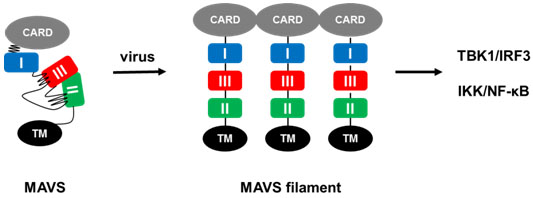
Fig. In unstimulated cells, MAVS is kept inactive by an autoinhibitory mechanism and upon viral infection, MAVS forms prion-like filaments to release its active regions for downstream signaling.
(Image provided by Prof. HOU Fajian`s group)
 Appendix: